

Here are pictures of the bacteria the characters are based on in The Rotters, taken using a scanning electron microscope to magnify the bacteria by up to 10,000 times. The accompanying captions (provided by the respective photographers) describe how the bacteria behave. (Click here for some child-friendly facts)
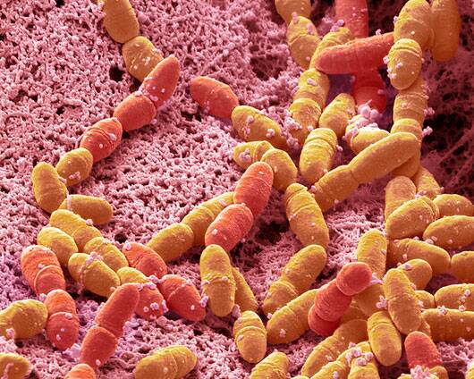
Streptococcus Mutans
(photographed by Steve Gschmeissner)
Here’s streptococcus mutans, as a coloured scanning electron micrograph (SEM). S. mutans is a coccoid shaped, Gram-positive, anaerobic bacteria that is part of the normal bacteria flora of the mouth. It metabolises sucrose to lactic acid and is a leading cause of tooth enamel decay. The acidic environment created in the mouth by this process is what causes the highly mineralized tooth enamel to decay. S. mutans is one of a few specialized organisms equipped with receptors for adhesion to the surface of teeth. Sucrose is utilized by S. mutans to produce a sticky, extracellular, dextran-based polysaccharide (glucan) that allows them to adhere to each other forming plaque. Other sugars (glucose, fructose, lactose) can be digested by S. mutans to produce lactic acid. It is the combination of plaque and acid that leads to dental decay. Magnification: x8000 when printed at 10cm wide.
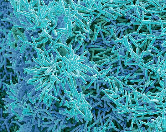
Plaque
(photographed by Steve Gschmeissner)
Plaque-forming bacteria, taken as a coloured scanning electron micrograph (SEM). Plaque consists of a film of bacteria embedded in a glycoprotein matrix. The matrix is formed from bacterial secretions and saliva. Plaque is the main cause of tooth decay. The bacteria feed on sugars in food, producing acid as a waste product. This acid corrodes the teeth’s enamel coating, resulting in dental caries. A build-up of dental plaque can also lead to inflamed and infected gums. Severe gum disease can lead to teeth falling out. Magnification: x3000 when printed at 10 centimetres across.
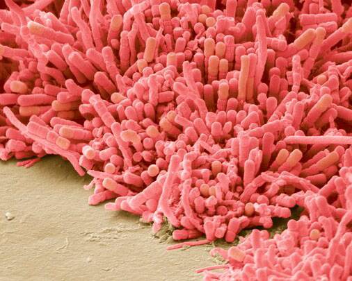
Plaque
(photographed by Steve Gschmeissner)
This is another close-up of plaque-forming bacteria, as a coloured scanning electron micrograph (SEM). The bacteria featured here are attached to the enamel of a child’s milk tooth. Magnification: x8000 when printed at 10 centimetres across.
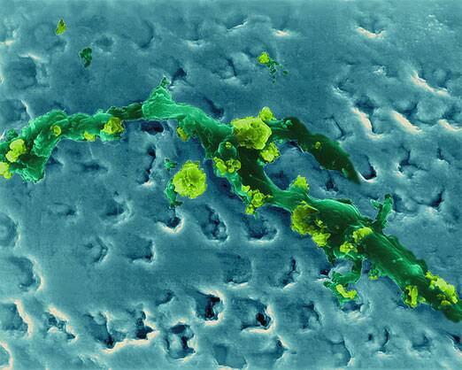
Tartar
(with plaque on it, photographed by Dennis Kunkel Microscopy)
This picture features a human tooth with an accumulation of bacterial plaque (smooth areas) and calcified tartar (rough areas) on the enamel surface, taken as a coloured scanning electron micrograph (SEM). Plaque is the sticky, colourless film of bacteria that forms on teeth. You can sense this film due to your tongue feeling a silky surface on your teeth. It is noticeable when teeth are not brushed. Plaque develops when foods containing carbohydrates (sugars and starches), such as milk, soft drinks or sweets are frequently left on the teeth. Bacteria that live in the mouth survive on these foods, producing acids as a result. Over time, these acids destroy tooth enamel, resulting in tooth decay. Plaque can also develop on the tooth roots under the gum and cause breakdown of the bone supporting the tooth. Tartar, or calculus, is plaque that has hardened on your teeth. Tartar can also form at and underneath the gum line and can irritate gum tissues. Magnification: x415 when shortest axis.